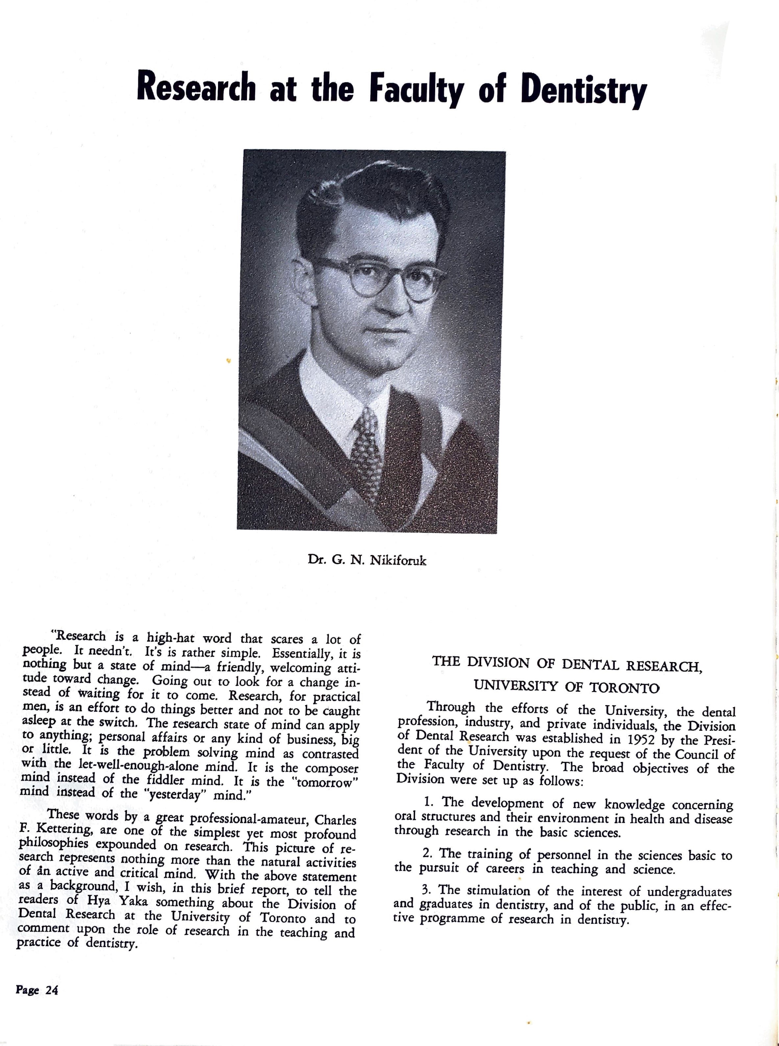
Dr. G. N. Nikiforuk
“Research is a high-hat word that scares a lot of people. It needn't. It's is rather simple. Essentially, it is nothing but a state of mind—a friendly, welcoming attitude toward change. Going out to look for a change instead of waiting for it to come. Research, for practical men, is an effort to do things better and not to be caught asleep at the switch. The research state of mind can apply to anything; personal affairs or any kind of business, big or little. It is the problem solving mind as contrasted with the let-well-enough-alone mind. It is the composer mind instead of the fiddler mind. It is the “tomorrow" mind instead of the “yesterday” mind.”
These words by a great professional-amateur, Charles F. Kettering, are one of the simplest yet most profound philosophies expounded on research. This picture of research represents nothing more than the natural activities of an active and critical mind. With the above statement as a background, I wish, in this brief report, to tell the readers of Hya Yaka something about the Division of Dental Research at the University of Toronto and to comment upon the role of research in the teaching and practice of dentistry.
THE DIVISION OF DENTAL RESEARCH,UNIVERSITY OF TORONTO
Through the efforts of the University, the dental profession, industry, and private individuals, the Division of Dental Research was established in 1952 by the President of the University upon the request of the Council of the Faculty of Dentistry. The broad objectives of the Division were set up as follows:
1. The development of new knowledge concerning oral structures and their environment in health and disease through research in the basic sciences.
2. The training of personnel in the sciences basic to the pursuit of careers in teaching and science.
3. The stimulation of the interest of undergraduates and graduates in dentistry, and of the public, in an effective programme of research in dentistiy.
these in one casing, and one no longer feared the firetouching the electric light installation. work display which could be caused by high voltage wire
The department in this school developed as an offshoot of the main clinic, but the records do not reveal the date when this occurred. One of the earliest names associated with X-rays in this school was Dr. F. Price, and another early worker in this field was Dr. R. D. Thornton. Later, radiology came under the direction of Dr. W. E. Willmott, a photograph of whose hands is displayed in the present intra oral room, and it was either during the time of Dr. Willmott or during the time of his successor, Dr. Richardson, that we were established as a separate department. Dr. Richardson remained as the Head of the department until 1945, and he was succeeded soon after by Dr. J. E. Moser, who held this position until his death in 1955. Dr. M. N. Rockman was then established in this appointment, which he held until 1957.
In the present department the oldest inhabitant is Dr. D. B. McAdam who was first appointed in 1953. For the past twelve years we have been fortunate in that Dr. Worth has travelled from Victoria every year in order to give the series of lectures on which the teaching of interpretation has been based.
In dental radiology at the present time, with one exception, we are in a period of modification of existing materials and equipment, for example, the improvement that has occurred in the quality of X-ray films over the past few years. The exception to which I referred is the increased awareness of the dangers associated with radiation. The knowledge that radiations are dangerous is not new, and there were papers on this subject as long ago as the early years of this century, and again about ten years ago there were some excellent articles on this theme. This aspect, however, has been brought into greater prominence recently because of two factors; firstly the increase and the potentially greater increase in general background radiation due to the introduction into the life of man of atomic and nuclear reactions, and secondly the greater realization of the implications of genetic changes in human beings which can be brought about by all radiations.
It is for this reason that in the Radiology Department today one hears so much talk of filtration of rays, limiting cones and changes in kilovoltage, and it is for this reason also that in the new department the walls of all the surgeries will be lined with lead.
The department at the new school will probably be as modern and as up-to-date as any that exists today, and it has been designed not only to cope with the present situation but with an anticipation of the future. There will be ten individual surgeries for intra oral radiography and one room planned for extra oral radiography in which will be housed apparatus for cephalometric surveys of a high degree of accuracy, and equipment for undertaking skull and general work. In addition to the normal requirements of radiography, there will be a teaching clinic room and a spare processing room to be used in cases of emergency, and for any special projects which may be undertaken.
It is always difficult to speculate on the future and it is no exception when speculating on possible trends and changes in this field, but, bearing in mind present research and the dental application of radiology in other fields, one can perhaps hazard a few guesses.
In the next few years we may see a still greater improvement in the X-ray films which are manufactured. Perhaps an emulsion will be produced for use with the higher kilovoltage apparatus, maintaining speed and at the same time giving greater contrast. This could facilitate interpretation of radiographs and enable the student to understand more fully the structures which he is viewing.
An adaptation of general medical radiology which we may see in dental use in the future is image intensification. This utilizes the principle of the television tube, and a weak image is increased in brightness by electronic activity.
An apparatus designed for dental work which has recently appeared on the market is the Rotagraph. This combines tomography with panoramic viewing and one is able to view on a flat surface a curved plane of a large mass. The practical application of this in dentistry is that theoretically one is able to view the whole of the mandible and maxilla on one film. In actual practice, this result is obtained by rotating the patient in one direction and the film in the opposite direction whilst a narrow beam of X-rays is directed at the patient. At some future date we may see such an apparatus in our new department.
Another adaptation which we may see in use in dentistry in the future is the point source of radiation from radio active material. This has been used in industry and general research for the detection of flaws and cracks in different materials, and in medicine it has been used experimentally. It has been suggested that it is not outside the bounds of possibility that a radio active material could be contained in the head of an instrument abou mensions of a pencil, and could be covered by a protective substance, except for one small opening. This could then be used either intra orally or extra orally with a film appropriately placed.
We have briefly passed through dental radiology and have finished in the realms of speculation. Perhaps we should get our feet on the ground again and consider the main aim of the department in the past, the present, and the future, and that is to equip the student to cope as efficiently as possible with all the problems in the field of dental radiology which he may encounter when in
practice.